Kissing spines on radiographs: what a finding says about pain and what it does not predict
Among 100 jumping horses in active competition, not one had a normal back on radiographs: what a kissing spines finding says, and what it cannot predict.
Scientific reviewPosition not yet filled, stated openly.
12 min readLast substantive review
Open access
Executive summary
A kissing spines finding describes close, touching or overlapping spinous processes on a radiograph. It is common in horses without back trouble: of 33 riding horses with no clinical signs, only seven were normal; of 100 jumping horses in competition, none. A higher grade is associated with back pain in horses presented for it, but the measurement depends on posture, and in one surgical series no imaging parameter predicted the outcome.
20primary sources
30 %of them level 1 to 2
2species studied
2004–2025publication years
Key points
Close or touching spinous processes are the rule in horses without perceived back pain: of 33 riding horses with no clinical signs, seven were entirely normal; of 100 jumping horses in active competition, none.
In 47 yearlings that had never been backed, about a third of the spaces were already narrowed or in contact, and their findings did not differ from those of 55 trained horses.
The finding is not empty for all that: among 582 horses presented for back pain or loss of performance, a higher radiographic grade went together with thoracolumbar pain, most strongly alongside changes in the small facet joints.
The narrowing that is measured depends on the exposure: manually induced flexion widened the space by up to 3.1 millimetres, against a threshold of four millimetres.
In the only series that tested it, neither the radiographic grade, nor the scintigraphic grade, nor the number of affected spaces predicted the return to performance after surgery.
What a kissing spines finding describes
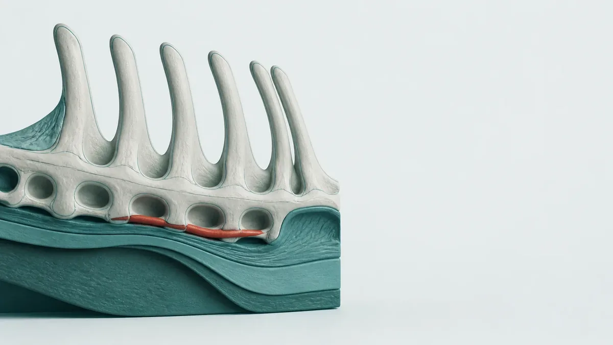
Along the back of every horse, the spinous processes of the thoracic and lumbar vertebrae rise like a row of bony sails. Kissing spines is the term used when neighbouring processes come very close, touch or overlap. The usual threshold is a space of less than four millimetres, with contact or overlap. The middle and caudal thoracic spine is most often affected, the lumbar region more rarely. The term therefore describes a shape of bone, not a state of the horse.
A 2025 review sums up the state of knowledge with unusual candour: how often pain really does arise from touching spinous processes remains speculative, no reliable agreement between the imaging finding and clinical signs could be shown, and the way the condition arises is poorly understood despite decades of treatments on offer. This is a narrative rather than a systematic review: it orders what is known, it does not measure it.
Is kissing spines a diagnosis?
No. The finding describes what can be seen on the radiograph. Whether a horse is in pain, and whether that pain comes from the spinous processes, is settled by the clinical examination, as a rule with diagnostic analgesia. Image and complaints are two separate pieces of information that then have to be brought together.
How common the finding is in horses without complaints
A Swedish study radiographed and scintigraphed 33 riding horses with no clinical signs of back problems. Only seven were entirely normal on both examinations, and eleven had a normal space of more than four millimetres throughout. Twenty-six showed at least one abnormality, usually mild and usually between the thirteenth and eighteenth thoracic vertebrae. The conclusion is blunt: the range of what is seen in horses without complaints is so wide that findings within that range cannot be read as clinically meaningful in horses that are unwell.
Twenty years later the same question was put to an entirely different population: 100 Warmblood jumping horses in active competition, average fence height 1.35 metres, with no observed lameness. Not one of these horses had a radiographically normal back. Narrowing and sclerosis lay mainly between the fifteenth thoracic and the first lumbar vertebra, and changes at the tendon attachments mainly in the region of the withers.
The most unexpected result of that work concerns the direction of the relationship: the highest career level ever reached was weakly but positively associated with narrowing of the spaces and with sclerosis. This must not be read as an advantage. The horses examined are those that lasted in the sport; those that failed do not appear in the sample, and that alone can produce such an association.
Where the changes come from is an open question. One study compared 47 yearlings that had never been backed with 55 trained Thoroughbreds without perceived back pain: the frequency, number and score of the findings did not differ. In the yearlings alone, about a third of the spaces examined were narrowed or in contact, and more than half showed sclerosis, lucencies or remodelling. The authors read this as a pointer towards a developmental origin. That reading is compatible with the data; the mere similarity of two groups does not prove it.
Why the finding is not meaningless all the same
The largest study on this question examined 582 horses presented for perceived back pain or loss of performance, all of which underwent diagnostic analgesia of the limbs, the back and the sacroiliac joints. Both the highest grade and the total grade of the spinous process changes were significantly associated with thoracolumbar pain, as was increased uptake on scintigraphy. Anyone who says the radiograph says nothing at all is contradicting this work.
Two qualifications from the same study weigh just as heavily. First, osteoarthritis of the small facet joints was more strongly associated with pain than the spinous processes alone, and the probability was highest when both were present together. Second, lameness of the fore or hind limbs and sacroiliac pain can mimic primary back pain, which is exactly why diagnostic analgesia remains indispensable. And because every horse had been referred with complaints, no figure for how common the finding is in the wider horse population follows from this work.
A French study compared 102 trotters with signs of back pain and 16 without. Ten of the 16 horses without complaints had changes, against 98 of the 102 symptomatic horses, though theirs were less severe and more localised. For simple contact between the spinous processes, however, the number of affected horses did not differ significantly between the groups: only the number of spaces involved and the severity did. The comparison group numbered 16 animals, which makes any conclusion about small differences uncertain.
A third study, in 223 Warmbloods, shows the same two-part pattern. Total grades on radiography and scintigraphy were higher in horses with thoracolumbar pain. The more finely calculated uptake ratios, by contrast, did not differ between horses with and without pain, and agreement between the scintigraphic methods and the radiographic finding was only slight.
How far the finding depends on the exposure itself
A radiograph is not a measurement but a projection. In six spinal columns from post-mortem examination, 49 spaces were measured 274 times and compared with cross-sectional images. In the backs classified as kissing spines, the spaces appeared clearly narrower on the radiograph than on the sagittal reconstruction. The projection therefore exaggerates the very narrowing on which the finding rests, and it does so under laboratory conditions: in the standing horse the geometry is harder to control, not easier.
The horse's posture shifts the result as well. In seven horses with already diagnosed contact, the thoracic spine was flexed by hand and radiographed in both postures. The space increased at every level from the fifth to the eighteenth thoracic vertebra, at most by a median of 3.1 and 3.0 millimetres, at least by 0.7 millimetres. As a reminder: the threshold below which a space counts as narrow is four millimetres.
A second, independent study points the same way: in seven healthy horses, traction on the abdomen widened the spaces from the eleventh thoracic to the second lumbar vertebra. Both studies cover seven horses; they show a direction, not a correction factor.
Observed shift in the measured width of the space when something about the exposure changes and nothing about the horse.
Condition changed
Effect on the measured width
Animals examined
Flexion of the thoracic spine by hand
Increase at every level from the fifth to the eighteenth thoracic vertebra, by a median of 0.7 to 3.1 millimetres
7 horses with diagnosed contact
Traction on the abdomen with a wide strap
Widening from the eleventh thoracic to the second lumbar vertebra, with a single exception; amount not quantified
7 healthy, sedated horses
Angle of the X-ray beam
Measured width altered, with no consistent pattern across the deviations
6 spinal columns from post-mortem examination
Projection instead of a cross-sectional image
Measured clearly narrower on radiography than on the sagittal reconstruction
6 columns classified as kissing spines
How consistently specialists read the same image
Images are read by people, and those people agree to varying degrees. In 100 healthy horses, three experienced assessors graded radiographs of the thoracic and lumbar spine independently of one another. Agreement was good for individual parameters: the width of the space, isolated sclerosis of the upper margin, beak-like new bone at the cranial dorsal aspect and remodelling at the cranial ventral aspect. It was only moderate for the total number of abnormalities per back, which is precisely the summary on which a purchase decision ultimately rests.
The jumping horse study likewise reports only moderate to good agreement between four assessors. Both figures come from study conditions, with experienced readers and standardised images. In everyday practice, between premises with different equipment and different training, rather less agreement is to be expected.
What does this mean for a pre-purchase examination?
There the radiographic finding supplies a description, not a prognosis. It says how the spinous processes look on that day, on that image. How reliably two specialists arrive at the same overall grade is only moderately documented, and none of the studies analysed here has tested whether this finding predicts the later course of a horse that is bought.
What the image predicts about the later course
The strongest objection to a large role for the radiograph comes from surgery itself. In a series of 71 horses in which the ligament between the spinous processes had been divided, 56 could be followed for a median of 35 months. Fifty-one of those 56 returned to some level of performance, a little more than nine in ten; 27 of 51 reached their previous level or a higher one.
What matters is what this study did not find. None of the variables tested influenced the return to performance: neither the number of affected spaces, nor the radiographic grade, nor the scintigraphic grade. The examination used to justify the procedure did not predict its outcome. This is the only study analysed here that expressly tested imaging grades as predictors, and 56 horses followed up is few: a weak association could be lost in a group of that size.
A second series, of 102 animals operated on standing, shows what counted instead. Of 66 horses with long-term follow-up, 53 returned to their intended work with no residual clinical signs. The difference lay not in the severity of the back finding but in whether another orthopaedic problem was present before surgery: without one, 28 of 30 horses returned; with one, 25 of 36. Thirty-six of the 102 are missing from the long-term follow-up, which can make the figures look kinder than they are.
The closest thing to a proper test of effectiveness is an analysis of racing performance. 159 operated Thoroughbred racehorses were compared with control horses matched for age, sex, race type, trainer and period, and followed for at least twelve months. The operated horses improved more than their controls. What was measured was racing performance, not pain, and allocation was clinical rather than random. Eight horses developed unilateral wasting of the back muscles with nerve involvement after the procedure.
The same question in humans
How much an imaging finding says about pain has been studied in human medicine for decades, and with study designs that equine medicine lacks. A systematic review of 33 studies covering 3,110 people without symptoms found signs of disc degeneration in 37 per cent of those in their twenties and in 96 per cent of those in their eighties. Bulges rose across the same age span from 30 to 84 per cent. Much of this, the authors conclude, belongs to normal ageing.
It does not follow that images mean nothing. A meta-analysis by the same group, covering 3,097 people up to 50 years of age, separated one sign from another: bulge, extrusion, certain signal changes and degeneration were markedly more common in people with low back pain than in controls without symptoms. Spinal stenosis, annular fissures and spondylolisthesis were not. The literature therefore does not say that the image is empty, but that the signs have to be examined one by one.
Two further studies concern the value of imaging itself. A meta-analysis of six randomised trials including 1,804 patients found that immediate imaging for low back pain without warning signs improved neither pain nor function. A systematic review of twelve follow-up studies found no consistent link between imaging findings and later low back pain; the predictive value is therefore unresolved rather than disproved.
One large randomised trial from health services research is particularly sobering. In four American health systems, imaging reports were supplemented with how often the finding in question occurs in people without symptoms; data from more than 238,000 adults were analysed. Use of health care services did not change measurably. Even supplying the ordinariness of a finding, then, barely shifts the decision.
The human counterpart to contact between the spinous processes even has a name of its own: Baastrup's disease. A 2022 systematic review of 35 studies and 1,308 patients describes it as a common and underdiagnosed cause of low back pain, whose recognition calls for imaging that includes functional views in flexion and extension. At the same time it records that the treatments applied rest on no firm basis and that randomised trials are lacking. On this one sign, then, human medicine is no further ahead than equine medicine.
What the radiograph can say and what it cannot
Taken together, the picture that emerges is neither reassuring nor alarming: it is blurred. Touching spinous processes are common in horses without complaints, they are already present in young animals that have never been ridden, and the narrowing that is measured depends on how the image came about. At the same time, in the largest study, a higher grade goes together with back pain.
Established: abnormalities of the spinous processes are the rule and not the exception in horses without perceived back pain.
Also established: higher grades go together with thoracolumbar pain in horses presented for it, especially alongside changes in the small facet joints.
Not established: that the grade of a radiographic finding tells you whether this particular horse is in pain.
Also not established: that the finding allows the course after surgery to be predicted; in the only series that tested it, it did not.
Not studied: how common touching spinous processes are in an unselected horse population. The percentages in circulation could not be traced in the specialist literature and are therefore not repeated here.
What remains useful in practice is the order of the questions. An image answers how the bone looks. Whether a horse is in pain and where that pain sits is answered by the clinical examination, by observation in movement and by diagnostic analgesia. Faced with a finding, most is gained by asking which of these steps were actually carried out, and what has changed since the horse last moved soundly.
What each study in this file contributes and which conclusion its design precisely does not support
Original analysis
Population, measured variable and central numerical result were taken from the original publication for every study used here; the last column names the conclusion that the study design does not support, and was derived specially for this overview.
Group examined
What was measured
Central result
Study design
What does not follow from it
33 riding horses with no back signs
Radiography and scintigraphy of the spinous processes
7 entirely normal, 11 with a normal space throughout
Cross-sectional, with no symptomatic comparison group
That abnormalities are harmless in horses that are unwell: the comparison group was missing
47 yearlings never backed against 55 trained Thoroughbreds
Radiography from the seventh thoracic to the third lumbar vertebra
No difference in frequency, number or score
Cross-sectional, a single breed
That training contributes nothing: equal values in two groups establish no cause
100 jumping horses in active competition
Radiography, four independent assessors
No horse without abnormalities; career level weakly associated with narrowing
Cross-sectional, in horses that lasted in the sport
That narrowing benefits performance: horses that failed are missing from the sample
102 trotters with and 16 without signs of back pain
Radiography from the fourteenth thoracic to the third lumbar vertebra
Changes in 10 of 16 horses without complaints and 98 of 102 symptomatic horses
Case-control study with a very small comparison group
That simple contact indicates pain: for that, the number of affected horses did not differ
582 horses at a referral clinic
Radiography, scintigraphy, diagnostic analgesia
Radiographic grade significantly associated with thoracolumbar pain
Cross-sectional, in cases preselected by complaints
A frequency figure for the wider horse population
6 spinal columns from post-mortem examination, 274 measurements
Radiography against cross-sectional imaging, different beam paths
Spaces measured clearly narrower on radiography than on cross-sectional images
Laboratory experiment under controlled conditions
That the discrepancy is just as large in the standing horse: there the geometry is harder to control
7 horses with diagnosed contact, two postures
Width of the space in a neutral and a flexed posture
Increase at every level, by a median of up to 3.1 millimetres
Case series, each horse serving as its own comparison
That flexion changes pain or the course of the condition: a distance was measured
100 healthy horses, three assessors
Agreement when grading
Good for individual parameters, only moderate for the total per back
Cross-sectional, under study conditions
That agreement is just as high in everyday practice
56 operated horses followed up
Return to performance after division of the ligament
A little more than nine in ten to some level, a little more than half to the previous level; no imaging parameter predicted this
Retrospective case series with no unoperated comparison group
That the procedure is the cause of the improvement
3,110 people without back pain
Magnetic resonance and cross-sectional images of the spine
Disc degeneration in 37 per cent of those in their twenties and 96 per cent of those in their eighties
Systematic review of 33 studies
A statement about spinous processes in the horse: another species, another structure, another technique
Limitations and uncertainty
There is no randomised study in the horse for any of the questions dealt with here. The editorial requirement to base at least half of the sources on the level of systematic review, meta-analysis or randomised trial cannot be met in this field: all six studies at that level come from human medicine, while the equine literature consists of cross-sectional studies, case-control studies and case series.
No study of how common touching spinous processes are in an unselected horse population exists. Every frequency figure comes from a selected group: riding horses at one clinic, jumping horses in competition, Thoroughbreds from one yard, trotters from one discipline.
In all the equine studies, the status of being free from back pain rests on clinical judgement, in part that of the people caring for the horses, and not on diagnostic analgesia. Some of the horses counted as unaffected may have been in pain.
The two studies on the influence of posture cover seven horses each, and the one on projection six spinal columns from post-mortem examination. They show a direction but supply no correction value for any one image.
The surgical series are retrospective and have no unoperated comparison group, and in both some of the horses are missing from the long-term follow-up. Neither proof of effectiveness nor a comparison between techniques follows from them.
The human work concerns intervertebral discs on magnetic resonance imaging for the most part. It supports the way of thinking about imaging findings, not the transfer of individual figures to the horse.
Open questions
How common are close or touching spinous processes in an unselected horse population, irrespective of the reason for presentation, the breed and the discipline?
Do the changes arise mainly during growth, as the comparison between yearlings and trained horses suggests, or does work alter them measurably over the years after all?
Can a standardised posture for the exposure be defined that makes measurement of the space comparable between practices and over time?
Is there any feature of the image or the examination that predicts the course after treatment, now that the imaging grades tested so far do not?
Frequently asked questions
My horse has kissing spines on the radiograph but shows no complaints. Is that serious?
The finding on its own says little about that. In a study of 33 riding horses with no back signs, only seven were entirely normal on radiography and scintigraphy, and among 100 jumping horses in active competition not one had a normal back. Abnormalities are therefore the rule in horses without complaints. At the same time, in the largest study, covering 582 horses presented for assessment, a higher grade went together with back pain. Both hold side by side, and the apparent contradiction is not resolved on the image but in the clinical examination of the individual horse.
Can a horse with kissing spines still be ridden?
That is settled neither by a radiograph nor by a study, but by veterinary examination of the individual horse. What the literature contributes is background: 100 jumping horses in active competition all had abnormalities of the spinous processes, and the highest career level reached was weakly but positively associated with narrowing. Those horses were working with the finding. No advantage can be read into that, because horses that failed in the sport are missing from such samples. Neither permission nor prohibition can be derived from the image.
Should I buy a horse with this finding?
There is no solid body of evidence on that, and this article gives no purchase recommendation. It is known that in 100 healthy horses three experienced assessors agreed well on individual parameters, but only moderately on the total number of abnormalities per back. Yet it is precisely that overall grade which carries a decision. It is also known that none of the studies analysed here has tested whether the finding predicts the later course of a horse that is bought. Anyone weighing it is weighing a description of bone, not a prognosis.
Why does one vet say narrow and the next one does not?
Because reading the image is less clear-cut than the word finding suggests. The width of a space is judged fairly consistently between experienced readers, whereas the overall assessment of a back is only moderately consistent. Added to that is the exposure itself: the angle of the beam alters the measured width, radiography measures narrower than a cross-sectional image, and flexion induced by hand widened the space by more than three millimetres in seven horses, against a threshold of four millimetres. Two images of the same horse are therefore not automatically comparable.
Does it come from riding or from a badly fitting saddle?
The evidence for that is missing, and one pointer argues against it. A study compared 47 yearlings that had never been backed with 55 trained Thoroughbreds without perceived back pain and found no difference in the frequency, number or score of the findings. In the yearlings alone, about a third of the spaces were narrowed or in contact. The authors read this as a pointer towards a developmental origin. That is not proof: equal values in two groups do not rule out an influence of work with any certainty. On the role of the saddle this study says nothing.
Do kissing spines occur in humans as well?
Yes, under the name Baastrup's disease, and the evidence there is no better. A 2022 systematic review of 35 studies and 1,308 patients describes contact between the spinous processes as a common and underdiagnosed cause of low back pain. At the same time it records that the treatments on offer rest on no firm basis and that randomised trials are lacking. The mean age was 59.6 years, which has no counterpart in the sport horse. What transfers is the way of thinking, not the figure.
Sources
Pilati N, Pressanto MC, Palumbo Piccionello A, De Angelis Corvi F. Impinging and Overriding Spinous Processes in Horses: A Narrative Review. Animals (Basel), 2025 (Other | Horse)DOI 10.3390/ani15182679 Narrative review: it records that the true frequency of pain caused by touching spinous processes remains speculative, that no agreement between the imaging finding and clinical signs could be shown, and that the way the condition arises remains poorly understood despite decades of treatments on offer.
Erichsen C, Eksell P, Holm KR, Lord P. Relationship between scintigraphic and radiographic evaluations of spinous processes in the thoracolumbar spine in riding horses without clinical signs of back problems. Equine Veterinary Journal, 2004 (Cross-sectional study | Horse)DOI 10.2746/0425164044877341 Of 33 riding horses with no clinical back signs, only 7 were normal on radiography and scintigraphy and 11 had a space of more than four millimetres throughout; the range seen in horses without complaints is, according to the authors, so wide that findings within it cannot count as clinically meaningful in horses that are unwell.
Pressanto MC, Pepe M, Coomer RPC, Pilati N. Radiographic abnormalities of the thoracolumbar spinous processes do not differ between yearling and trained Thoroughbred horses without perceived back pain. Journal of the American Veterinary Medical Association, 2023 (Cross-sectional study | Horse)DOI 10.2460/javma.22.09.0419 Between 47 yearlings that had never been backed and 55 trained Thoroughbreds without perceived back pain, the frequency, number and score of the findings did not differ, while in the yearlings alone about a third of the spaces were already narrowed or in contact: the authors read this as a pointer towards a developmental origin.
Kerbert MP, le Jeune SS, Philips KL, Shaw KL. Mild radiographic abnormalities of thoracolumbar spinous processes are not associated with decreased performance in 100 Warmblood jumping horses in active competition. Journal of Equine Veterinary Science, 2025 (Cross-sectional study | Horse)DOI 10.1016/j.jevs.2025.105366 In 100 jumping horses in active competition not one back was radiographically normal, and the highest career level reached was weakly but positively associated with narrowing and sclerosis: a result the authors read as a sign that mild findings do not necessarily impair performance.
Looijen MGP, Hanousek K, Maree JTM, Visser MC. Inter- and intra-observer agreement for grading radiographs of thoracolumbar spinous processes in healthy horses during a pre-purchase examination. The Veterinary Record, 2022 (Cross-sectional study | Horse)DOI 10.1002/vetr.1065 Three assessors agreed well on individual parameters such as the width of the space, but only moderately on the total number of abnormalities per back, which is why the authors call for only reliably readable parameters to be used for grading in future.
Cousty M, Retureau C, Tricaud C, Geffroy O. Location of radiological lesions of the thoracolumbar column in French trotters with and without signs of back pain. The Veterinary Record, 2010 (Case-control study | Horse)DOI 10.1136/vr.c70 In 102 trotters with and 16 without signs of back pain, the lesions in the horses without complaints were less severe and more localised, but for simple contact between the spinous processes the number of affected horses did not differ significantly between the groups.
Zimmerman M, Dyson S, Murray R. Close, impinging and overriding spinous processes in the thoracolumbar spine: the relationship between radiological and scintigraphic findings and clinical signs. Equine Veterinary Journal, 2012 (Cross-sectional study | Horse)DOI 10.1111/j.2042-3306.2011.00373.x In 582 horses presented for back pain or loss of performance there was a significant link between the radiographic grade and thoracolumbar pain, but limb lameness and sacroiliac joint pain can mimic primary back pain, which is why diagnostic analgesia remains indispensable.
van Zadelhoff C, Ehrle A, Merle R, Jahn W. Thoracic processi spinosi findings agree among subjective, semiquantitative, and modified semiquantitative scintigraphic image evaluation methods and partially agree with clinical findings in horses with and without thoracolumbar pain. Veterinary Radiology & Ultrasound, 2019 (Cross-sectional study | Horse)DOI 10.1111/vru.12695 In 223 Warmbloods the total grades on radiography and scintigraphy were higher in horses with thoracolumbar pain, while the semiquantitative uptake ratios did not differ and agreement between the scintigraphic methods and the radiographic finding was only slight.
Djernaes JD, Nielsen JV, Berg LC. Effects of X-ray beam angle and geometric distortion on width of equine thoracolumbar interspinous spaces using radiography and computed tomography-a cadaveric study. Veterinary Radiology & Ultrasound, 2017 (Laboratory study | Horse)DOI 10.1111/vru.12466 In six spinal columns from post-mortem examination, beam path and geometric distortion altered the measured width of the spaces, and in backs classified as kissing spines the radiographic measurement was clearly narrower than on the sagittal cross-sectional reconstruction.
O'Sullivan S, McGowan CM, Junnila J, Hyytiäinen HK. The effect of manually facilitated flexion of the thoracic spine on the interspinous space among horses with impinging dorsal spinous processes of the thoracic vertebrae. The Veterinary Journal, 2022 (Case series | Horse)DOI 10.1016/j.tvjl.2022.105909 In seven horses with diagnosed contact, flexion induced by hand widened the space at every level from the fifth to the eighteenth thoracic vertebra, by a median of 0.7 to 3.1 millimetres, which according to the authors can change the grade assigned and the decision that follows from it.
Pigé C, Masseau I, Bonilla AG. Influence of abdominal elevation on radiographic measurements of the thoracolumbar interspinous spaces in asymptomatic horses. The Canadian Veterinary Journal, 2020 (Other | Horse)PMID 33012827 In seven horses without complaints, traction on the abdomen widened the spaces from the eleventh thoracic to the second lumbar vertebra with a single exception; the work was intended as a surgical aid and shows independently that posture at the moment of exposure helps determine the measurement.
Prisk AJ, García-López JM. Long-term prognosis for return to athletic function after interspinous ligament desmotomy for treatment of impinging and overriding dorsal spinous processes in horses: 71 cases (2012-2017). Veterinary Surgery, 2019 (Case series | Horse)DOI 10.1111/vsu.13298 Of 56 operated horses followed up, 51 returned to some level of performance and 27 of 51 to their previous level or a higher one, with neither the number of affected spaces nor the radiographic or scintigraphic grade influencing that return.
de Souza TC, Crowe OM, Bowles D, Poore LA. Minimally invasive cranial ostectomy for the treatment of impinging dorsal spinous processes in 102 standing horses. Veterinary Surgery, 2022 (Case series | Horse)DOI 10.1111/vsu.13736 Of 66 horses with long-term follow-up, 53 returned to their intended work with no residual clinical signs, the difference lying not in the severity of the back finding but in whether another orthopaedic problem was present before surgery (28 of 30 against 25 of 36).
Derham AM, O'Leary JM, Connolly SE, Schumacher J. Performance comparison of 159 Thoroughbred racehorses and matched cohorts before and after desmotomy of the interspinous ligament. The Veterinary Journal, 2019 (Cohort study | Horse)DOI 10.1016/j.tvjl.2019.05.004 159 operated Thoroughbred racehorses improved significantly more than comparison horses matched for age, sex, race type, trainer and period, while eight horses developed unilateral wasting of the back muscles with nerve involvement and allocation was clinical rather than random.
Brinjikji W, Luetmer PH, Comstock B, Bresnahan BW. Systematic literature review of imaging features of spinal degeneration in asymptomatic populations. AJNR American Journal of Neuroradiology, 2015 (Systematic review | Human)DOI 10.3174/ajnr.A4173 In 33 studies covering 3,110 people without symptoms, disc degeneration rose from 37 per cent in those in their twenties to 96 per cent in those in their eighties, which leads the authors to assign many of these signs to normal ageing and to call for interpretation only in the clinical context.
Brinjikji W, Diehn FE, Jarvik JG, Carr CM. MRI Findings of Disc Degeneration are More Prevalent in Adults with Low Back Pain than in Asymptomatic Controls: A Systematic Review and Meta-Analysis. AJNR American Journal of Neuroradiology, 2015 (Meta-analysis | Human)DOI 10.3174/ajnr.A4498 In 14 studies covering 3,097 people up to 50 years of age, bulge, extrusion, degeneration and certain signal changes were more common in people with low back pain, whereas spinal stenosis, annular fissures and spondylolisthesis were not: imaging findings have to be examined one by one.
Chou R, Fu R, Carrino JA, Deyo RA. Imaging strategies for low-back pain: systematic review and meta-analysis. The Lancet, 2009 (Meta-analysis | Human)DOI 10.1016/S0140-6736(09)60172-0 In six randomised trials including 1,804 patients, immediate imaging for low back pain without warning signs improved neither pain nor function, neither in the short term nor after six to twelve months.
Steffens D, Hancock MJ, Maher CG, Williams C. Does magnetic resonance imaging predict future low back pain? A systematic review. European Journal of Pain, 2014 (Systematic review | Human)DOI 10.1002/j.1532-2149.2013.00427.x In twelve follow-up studies no consistent link emerged between magnetic resonance findings and later low back pain, but inconsistency and low quality prevented any pooling: the predictive value is unresolved rather than disproved.
Jarvik JG, Meier EN, James KT, Gold LS. The Effect of Including Benchmark Prevalence Data of Common Imaging Findings in Spine Image Reports on Health Care Utilization Among Adults Undergoing Spine Imaging: A Stepped-Wedge Randomized Clinical Trial. JAMA Network Open, 2020 (Randomised trial | Human)DOI 10.1001/jamanetworkopen.2020.15713 Among more than 238,000 adults analysed, stating in the report how common a finding is in people without symptoms did not measurably change the use of health care services, but slightly reduced the prescribing of opioids.
Meluzio MC, Smakaj A, Perna A, Velluto C. Epidemiology, diagnosis and management of Baastrup's disease: a systematic review. Journal of Neurosurgical Sciences, 2022 (Systematic review | Human)DOI 10.23736/S0390-5616.21.05428-X 35 studies covering 1,308 patients describe the human counterpart, Baastrup's disease, as a common and underdiagnosed cause of low back pain, while recording at the same time that the conservative and surgical treatments applied rest on no firm basis and that randomised trials are lacking.
ForschungPferd (2026). Kissing spines on radiographs: what a finding says about pain and what it does not predict. ForschungPferd, English. https://forschungpferd.ch/en/equine-health/kissing-spines-radiographic-findings/