Scientific reviewPosition not yet filled, stated openly.
11 min readLast substantive review
Open access
Executive summary
Laminitis announces itself through no single reliable sign. In British practice data, bilateral forelimb lameness separated laminitis from other lameness more sharply than any other sign, followed by an increased digital pulse; together the two covered 99 cases in 100. These figures come from a comparison with horses that were already lame. How early a sign appears has not so far been quantified by any study.
22primary sources
23 %of them level 1 to 2
1species studied
1998–2025publication years
Key points
In a British analysis of 588 veterinary confirmed laminitis cases set against 201 horses lame for another reason, bilateral forelimb lameness was the single strongest sign: 92 of every 100 horses showing it had laminitis.
Together with an increased digital pulse, this pair of signs captured 99 per cent of the laminitis cases in that survey. Both figures apply to a comparison among lame horses, not to a horse that moves normally.
Around 45 per cent of the veterinary confirmed cases had not been recognised as laminitis by their keepers; the call concerned undefined lameness, a suspected hoof abscess, colic or stiffness.
The earliest measurable signal does not lie in the hoof: among 374 ponies that had never been affected, the highest band of fasting insulin went with 69 per cent developing laminitis within four years, against 6 per cent in the lowest band.
No study found in this search puts a figure on the time between the first visible sign and damage to the suspensory apparatus.
What happens in laminitis, and how often it occurs
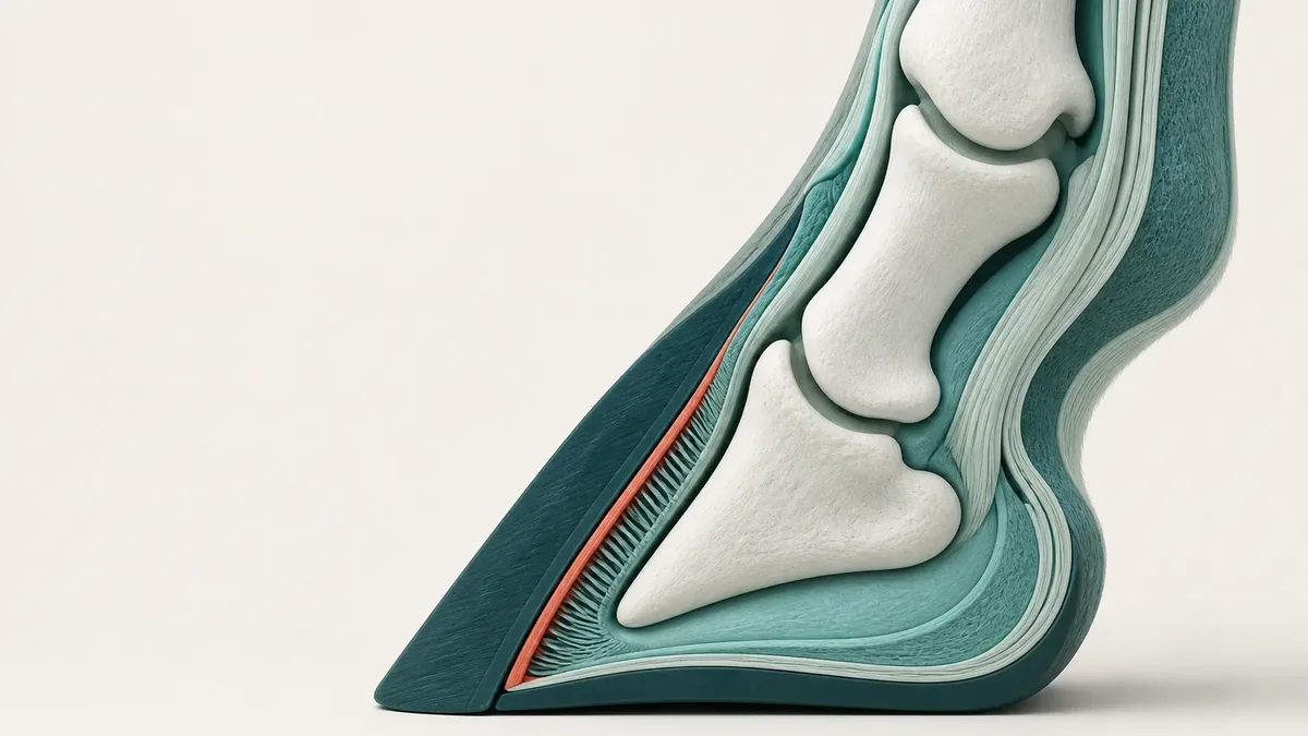
Laminitis does not affect the whole hoof but a narrow zone inside it: the interlocking lamellae that suspend the pedal bone from the inner hoof wall. When that attachment fails, the bone shifts against the wall. Two consequences follow: the disease usually affects several hooves at once, and it is severely painful, because the tissue involved is load-bearing and under full weight.
How common laminitis is proves surprisingly hard to establish. A systematic review screened 69 publications and kept 10 as usable. The reported frequencies ranged from 1.5 to 34 per cent, and the authors state that comparing those figures with one another would be illegitimate, because they concern different causes and populations. That range is therefore not a prevalence but the admission of a gap.
More robust is a prospective British cohort of 1,070 horses and ponies: 123 episodes in 97 animals over a little more than two years. A first episode occurred at a rate of around 9.6 cases per 100 horse years, with 11.5 for recurrences. One secondary result deserves attention: barely half of the reported episodes were diagnosed by a vet at all. Analyses that count only veterinary cases therefore underestimate the true frequency in a systematic way.
Which signs separate laminitis most sharply from other lameness
The only work that weighs early signs against one another comes from British first-opinion practice. It compares 588 laminitis cases diagnosed by a vet with 201 horses that were lame for another reason. Five signs differed between the groups by more than 50 percentage points in frequency; for others, the work gives the strength of the link with laminitis.
Bilateral forelimb lameness separated most strongly: in horses showing it the odds of laminitis were around 40 times higher, and 92 in 100 of them did have laminitis. An increased digital pulse followed, with odds around 13 times higher. Taken together, the two signs captured 99 per cent of the laminitis cases in that survey.
Clinical signs compared between 588 laminitis cases and 201 horses lame for another reason, ordered by the strength of the link.
Sign
Odds of laminitis against horses lame for another reason
Bilateral forelimb lameness
around 40-fold
Shifting weight from limb to limb
around 18-fold
Difficulty turning
around 17-fold
Flat or convex sole
around 15-fold
Increased digital pulse
around 13-fold
Short, stiff gait at the walk
around 9-fold
Reluctance to move forward
around 4-fold
Can I use this to check for laminitis myself?
No. All the comparison horses in that study were already lame, and the signs were ticked off a list by vets. The figures therefore say how well a sign separates laminitis from other causes of lameness, and precisely not what it means in a horse that moves normally. The authors expressly call their work a first evidence base, not a validated test.
The digital pulse: physiologically supported, barely tested
The digital pulse is the pulsation of the arteries running down past the fetlock towards the hoof. That a pulse felt as increased really does reflect something is established. In 42 horses examined by Doppler, blood flow in the lateral digital artery was markedly higher, in a systemic inflammatory response, when the pulse was palpably increased. In horses that already had laminitis the artery was also wider.
The experiment shows the sign as well. In a randomised model in which a high insulin level was maintained for 48 hours in young Standardbred horses, all four treated animals developed laminitis with marked digital pulses; the four control animals remained unremarkable.
How reliable is the assessment of such a sign?
This is the open flank of the subject. For the digital pulse itself, no study of agreement between examiners was found in this search. Only the severity scale for laminitic lameness has been tested: the same vet agreed with herself only moderately, and different vets with one another somewhat better.
Those figures come from a study in which 25 ponies were assessed twice and 13 video recordings were then graded independently by 58 experienced equine vets. Collapsing the four severity grades into three levels, sound, mild and severe, improved agreement markedly. If the established scale is only moderately reproducible, no greater accuracy should be expected from a palpated pulse.
Hoof warmth: two studies, two directions
Warm hooves count as a classic early sign, and keepers report them more often than vets do. Hoof temperature has rarely been measured, however, and the two studies that did so point in different directions.
In a feeding model from 1998, 13 of 21 Standardbred horses received a starch overload. Between the twelfth and the thirty-second hour afterwards, hoof temperature was the only measurement that separated the animals that later fell ill from the rest. The finding is real, but its reach is narrow: it came from a continuous temperature recorder, in a chamber cooled to ten degrees, and therefore against a background of cold-induced vasoconstriction. It says nothing about a hand laid on the hoof in the stable.
The only work found on the diagnostic value of thermal imaging in the lame horse reaches the opposite result. Among 36 sound horses and 119 lame ones, navicular disease, inflammation of the hoof corium and tendon disease differed clearly from the sound animals, but laminitis of all things did not. The likeliest explanation lies in the method: what was measured was the temperature difference between the left and the right forelimb. A horse affected in both forelimbs stays symmetrical in that comparison, and so invisible.
What is actually seen at the yard
How well early signs register in daily life was studied in 93 veterinary confirmed cases from 25 British practices. The result has two sides. Every suspicion voiced by a keeper was confirmed: 51 out of 51. At the same time 42 of the 93 cases, a little over 45 per cent, had not been recognised as laminitis. The call then concerned undefined lameness, a suspected hoof abscess, colic or stiffness.
Who notices what is instructive too. Keepers reported warm hooves more often, vets more often reported difficulty turning, weight shifting and body condition. In the recognised cases, divergent hoof rings and breed type were documented markedly better than in the rest. Anyone who already saw a horse at risk read the same signs differently.
How wide the gap can grow shows in 38 horses with confirmed disease of the pituitary gland. Owners reported laminitis in 37 per cent, radiography found it in 76 per cent. Recognition rose with the severity of the insulin elevation: the quiet forms slip through exactly where they are most common.
In the prospective cohort of 1,070 animals the most frequently reported signs were neither pulse nor warmth but two features of movement: difficulty turning and a short, stiff or lame gait, each in more than 70 per cent of episodes. Both forelimbs were affected in around 63 per cent of episodes, and three quarters of the animals had had laminitis before.
Why ‘early’ is a question without a solid answer here
The most obvious question is how much time lies between the first visible sign and damage to the suspensory apparatus. The literature gives no answer: no study putting a figure on that interval was found here. What circulates comes from experimental models, and those measure something else.
In nine sound ponies given insulin over a prolonged period, second grade laminitis appeared in all four hooves after around 55 hours on average. That is the time from the start of the insulin load, not from the first sign, and the level maintained was several times the animals' own baseline.
A tissue study in the same model goes further still. Six hours after the start of the load the secondary lamellae were already narrowed and basal cells had died, at a stage when nothing was yet visible clinically. Inflammatory cells and damage to the basement membrane appeared only after 24 to 48 hours, which the authors classify as a later event rather than the initial cause. The damage precedes the sign.
Natural cases point the same way. In 14 laminitic horses with raised insulin, the severity of the tissue changes bore no relation to the duration of illness reported by the owner and must, at least in part, have preceded it. The authors regard repeated, unnoticed episodes as the most plausible explanation.
A Swiss study fits this picture. In 51 elderly horses at the Bern equine clinic, clinically unremarkable and not lame, radiography revealed signs of chronic laminitis. Age over 25 years went with it, but neither body condition, nor the crest of the neck, nor the pituitary hormone value. A horse nobody takes to be laminitic may already carry the marks.
The earliest measurable signal is not in the hoof
Anyone who wants to be genuinely early must look before the hoof. In a cohort of 374 ponies that had never had laminitis, followed every six months for up to four years, two blood values quantified future risk best: fasting insulin and insulin one hour after sugar given by mouth. In the lowest fasting group, which held 70 per cent of the ponies, 6 per cent fell ill within four years, against 22 per cent in the middle fifth and 69 per cent in the top tenth.
A second experiment supports the direction. 37 ponies received a starch-rich ration after a sugar load by mouth. In the animals with the lowest insulin response no laminitis occurred; in the middle band it affected a good third, and in the highest six ponies out of seven.
What does not carry in either study is notable. A raised value of the pituitary hormone ACTH without clinical signs of pituitary disease was only weakly linked to laminitis, and in the pony cohort that hormone was not independently associated with risk. The common picture, in which this hormonal disorder explains laminitis by itself, falls short.
A post-mortem finding fits: in 16 horses with confirmed pituitary disease, the lamellae of the ten without laminitis were microscopically normal, and all six laminitic animals had raised insulin. In a Finnish case series of 36 laminitic horses, around 89 per cent had an underlying hormonal disease. How much an established insulin disorder weighs shows finally in the control arm of one trial: on a starch-rich ration, 14 of 37 untreated ponies developed laminitis.
Risk factors, season and the route to diagnosis
The familiar story of spring grazing holds up only partly against the data. In a case-control study with 191 laminitis cases and 819 comparison horses, the summer and winter months were linked to raised risk relative to spring. Added to these were weight gain in the previous three months, new access to grazing in the last four weeks, box rest in the preceding week, previous laminitis, sensitivity after shoeing or trimming, a known hormonal disease, and a growing interval since the last worming. The prospective cohort, for its part, found no meaningful difference in frequency between the seasons.
Some results from the same study run against expectation: increasing height, supplementary feeds and recent transport went with lower risk, without any mechanistic explanation. Findings of that kind are a good reason to read case-control data as links rather than as causes.
How weakly the field stands overall is recorded by a systematic review: 17 fully appraised publications, 6 of them carrying the most reliable information, and an overall picture the authors call inconsistent. Only the link between increasing age and chronic laminitis is well supported; sex, breed and weight remained contradictory.
Diagnosis is secured by veterinary examination and radiography. A 2025 study of 126 radiographs from 50 horses shows that the lucent zone between inner hoof wall and pedal bone separates acute cases from sound horses usefully, and clearly better than the classic measurements along the outer wall. That measurement too was taken in horses already regarded as laminitic: it confirms and grades, it does not search.
Test bench for the early signs of laminitis: what has been tested and what stays open
Original analysis
For each sign the methodologically strongest study design found in this search is entered, together with the group it was compared against and, in the last column, the test that is still missing for that sign.
Sign or measurement
Best design found
Compared against
What it supports
What has not been tested
Bilateral forelimb lameness
Cross-sectional study of 588 cases
Horses lame for another reason
The single strongest sign in that survey
Its meaning in a horse that is not lame; agreement between examiners
Increased digital pulse
Cross-sectional study, plus Doppler measurement and a randomised insulin model
Horses lame for other reasons and untreated control animals
That the palpation finding reflects a real change in blood flow
Agreement between two people palpating the same pulse
Warm hooves
Feeding model with continuous temperature recording, plus a thermal imaging study
Experimental animals that stayed healthy and sound horses
A rise in temperature in the model between the twelfth and the thirty-second hour
The value of a hand in the stable; thermal imaging found no difference in laminitis
Difficulty turning
Cross-sectional study and prospective cohort
Horses lame for another reason
The sign keepers report most often
Whether it appears earlier than the other signs
Divergent hoof rings
Cross-sectional study of 93 cases, also included in the pony cohort model
Cases not recognised as laminitis
More often documented where keepers did recognise the laminitis
Whether they indicate a current episode or a past one
Fasting insulin
Prospective cohort over four years
Ponies of the same cohort in lower bands
A strong grading of risk over several years
Transfer to other measurement methods, breeds and regions
Insulin after sugar given by mouth
Prospective cohort and feeding experiment
Ponies with a lower insulin response
A grading of risk before anything is visible at the hoof
Its reach beyond the pony populations studied
Lucent zone on radiography
Retrospective analysis of 126 radiographs
Sound horses from the same clinic
Separation of acute cases from sound horses
Its suitability as a screening test in horses without signs
Limitations and uncertainty
The editorial requirement that at least half the sources sit at the level of systematic review, meta-analysis or randomised trial cannot be met here: only five of the studies used reach that level. The field consists mainly of cross-sectional, case-control and cohort studies, together with induction models in small numbers of animals.
The weighting of the clinical signs comes from a single cross-sectional study that has never been confirmed externally. Its comparison group consisted only of lame horses, the signs were ticked without any check on agreement between examiners, and the publication names only signs above a threshold of 50 percentage points. The absence of a sign from that list does not mean it was not recorded.
For the digital pulse and for hoof warmth, no study of agreement between different examiners was found. Both count as classic early signs, yet their assessment in the living horse has never been tested for reproducibility.
The insulin values come from pony cohorts and from feeding experiments with a set ration. The authors expressly state that the figures do not apply to other measurement methods, other breeds, other regions or other management systems.
None of the studies found puts a figure on the interval between the first visible sign and measurable damage. The hours often quoted come from induction models and measure the time from the start of an artificially produced insulin load.
The field and practice data come almost entirely from Great Britain, the models from Australia and Finland. The one Swiss study covers 51 elderly horses, defines laminitis purely on radiography and did not analyse insulin.
Open questions
How well do two experienced people agree when they palpate the same digital pulse, and how well does the same person agree with herself?
How much time passes between the first sign visible on the horse and measurable damage to the lamellae?
How common are repeated, unnoticed episodes, and can they be demonstrated in the living horse rather than only at post-mortem?
Do the insulin bands derived in ponies keep their meaning in larger horses and under central European management conditions?
Frequently asked questions
My horse has warm hooves. Is that laminitis already?
Warm hooves on their own support no diagnosis. Hoof temperature varies a great deal in daily life, and the only thermal imaging study in the lame horse found, for laminitis of all conditions, no meaningful difference from sound animals, very probably because it measured the difference between the left and the right forelimb, in which bilateral disease stays symmetrical. In a feeding model hoof temperature was indeed the only separating measurement between the twelfth and the thirty-second hour, but it was taken with a continuous recorder in a cooled chamber. If warmth comes with a short, stiff way of moving or with forelimb lameness, that belongs with the veterinary practice.
Is a strong digital pulse a reliable sign of laminitis?
Reliable it is not, but it is among the strongest. In the British practice study the odds of laminitis with an increased digital pulse were around 13 times higher than in horses lame for another reason, and together with bilateral forelimb lameness it captured 99 per cent of the cases in that survey. A Doppler measurement confirms that a palpably increased pulse really does go with higher blood flow. The decisive question stays open: how alike two people judge the same pulse has not been published so far. An increased pulse also occurs in other diseases of the hoof.
How can I recognise laminitis as early as possible?
On present data it cannot be recognised early in any reliable way, and that is the most honest answer. In the prospective cohort the signs reported most often were difficulty turning and a short, stiff or lame gait, each in more than 70 per cent of episodes. At the same time, in a practice study a little over 45 per cent of confirmed cases went unrecognised by their keepers. Tissue studies show that the damage precedes the visible sign. Anyone who wants to judge risk genuinely early cannot avoid metabolism: the insulin values graded risk years in advance.
Does laminitis really occur mainly in spring?
No, and the data contradict that picture quite clearly. In the prospective cohort of 1,070 horses and ponies the authors found no meaningful difference in frequency between the seasons. The case-control study with 191 cases even found raised risk in the summer and winter months relative to spring. What stood out in that same study was less the season than change: weight gain in the previous three months, new access to grazing in the last four weeks and box rest in the preceding week.
Does laminitis always affect the front hooves?
No. In the prospective cohort both front hooves were affected in around 63 per cent of episodes, so other distributions occurred in the rest. How those remaining episodes break down in detail the analysis does not say, which is why no proportion for purely hind involvement can be given here. In practice this means two things: the typical bilateral forelimb lameness is the most common presentation but not the only one, and an unusual distribution does not rule laminitis out. For judging an individual horse, the examination on site is what counts.
My horse is not overweight. Can it still get laminitis?
Yes. The systematic review of risk factors expressly calls the findings on weight, breed and sex contradictory; only the link with increasing age in chronic laminitis is well supported. In the Swiss study of 51 elderly horses, neither body condition nor the assessment of the crest of the neck was linked to radiographic signs of laminitis, whereas age over 25 years was. Conversely, in a Finnish case series almost all laminitic horses with raised insulin showed outward signs of being overweight. What appears to matter is the insulin situation, not the shape alone.
Sources
Wylie CE, Shaw DJ, Verheyen KLP, Newton JR. Decision-tree analysis of clinical data to aid diagnostic reasoning for equine laminitis: a cross-sectional study.. Veterinary Record, 2016 (Cross-sectional study | Horse)DOI 10.1136/vr.103588 In the comparison of 588 veterinary diagnosed laminitis cases with 201 horses lame for another reason, bilateral forelimb lameness was the single strongest sign and the increased digital pulse the second strongest; the authors expressly describe their work as a first evidence base for assessing clinical signs of laminitis, not as a validated test.
Pollard D, Wylie CE, Verheyen KLP, Newton JR. Assessment of horse owners' ability to recognise equine laminitis: A cross-sectional study of 93 veterinary diagnosed cases in Great Britain.. Equine Veterinary Journal, 2017 (Cross-sectional study | Horse)DOI 10.1111/evj.12704 All 51 suspicions of laminitis voiced by keepers were confirmed by the vet, yet 42 of the 93 cases had not been recognised as laminitis; warm hooves were reported more often by keepers, difficulty turning and weight shifting more often by vets.
Pollard D, Wylie CE, Newton JR, Verheyen KLP. Incidence and clinical signs of owner-reported equine laminitis in a cohort of horses and ponies in Great Britain.. Equine Veterinary Journal, 2019 (Cohort study | Horse)DOI 10.1111/evj.13059 In a prospective cohort of 1,070 animals with 123 episodes there was no meaningful difference in frequency between the seasons, and barely half of the episodes were diagnosed by a vet, which is why analyses of veterinary cases alone underestimate the frequency.
Wylie CE, Collins SN, Verheyen KLP, Newton JR. Risk factors for equine laminitis: a case-control study conducted in veterinary-registered horses and ponies in Great Britain between 2009 and 2011.. The Veterinary Journal, 2013 (Case-control study | Horse)DOI 10.1016/j.tvjl.2013.08.028 With 191 cases and 819 comparison horses, the summer and winter months were linked to raised risk relative to spring, as were recent weight gain, new access to grazing, box rest, previous laminitis, sensitivity after shoeing or trimming and a known hormonal disease.
Wylie CE, Collins SN, Verheyen KLP, Newton JR. Risk factors for equine laminitis: a systematic review with quality appraisal of published evidence.. The Veterinary Journal, 2012 (Systematic review | Horse)DOI 10.1016/j.tvjl.2011.10.020 Of 17 fully appraised publications, 6 counted as the most reliable source of information; the authors describe the epidemiological findings on spontaneous laminitis as inconsistent and regard only the link between increasing age and chronic laminitis as well supported.
Wylie CE, Collins SN, Verheyen KLP, Newton JR. Frequency of equine laminitis: a systematic review with quality appraisal of published evidence.. The Veterinary Journal, 2011 (Systematic review | Horse)DOI 10.1016/j.tvjl.2011.04.014 Of 69 appraised publications, 10 provided the most reliable information; the reported frequencies of 1.5 to 34 per cent must not, according to the authors, be compared with one another because they concern different causes, so the true frequency remains unknown.
Menzies-Gow NJ, Stevens KB, Sepulveda MF, Jarvis N, Marr CM. Repeatability and reproducibility of the Obel grading system for equine laminitis.. Veterinary Record, 2010 (Cross-sectional study | Horse)DOI 10.1136/vr.c3668 With 25 ponies and 58 assessing vets the established severity scale reached only moderate agreement of one person with herself and substantial agreement between people; collapsing the grades into sound, mild and severe improved agreement markedly.
Aguirre CN, Talavera J, Fernández Del Palacio MJ. Usefulness of Doppler ultrasonography to assess digital vascular dynamics in horses with systemic inflammatory response syndrome or laminitis.. Journal of the American Veterinary Medical Association, 2013 (Cross-sectional study | Horse)DOI 10.2460/javma.243.12.1756 In 42 horses, blood flow in the lateral digital artery was markedly higher when the digital pulse was palpably increased, which supports the palpation finding physiologically; the authors see the Doppler measurement as a complementary method and not as a replacement for palpation.
de Laat MA, McGowan CM, Sillence MN, Pollitt CC. Equine laminitis: induced by 48 h hyperinsulinaemia in Standardbred horses.. Equine Veterinary Journal, 2010 (Randomised trial | Horse)DOI 10.2746/042516409X475779 A high insulin level alone triggered laminitis within 48 hours in all four treated Standardbred horses, with marked digital pulses against unremarkable controls; wall temperature was higher but varied less than in the control animals.
Asplin KE, Sillence MN, Pollitt CC, McGowan CM. Induction of laminitis by prolonged hyperinsulinaemia in clinically normal ponies.. The Veterinary Journal, 2007 (Randomised trial | Horse)DOI 10.1016/j.tvjl.2007.07.003 All five treated ponies developed laminitis in all four hooves after around 55 hours of artificially raised insulin, and no control animal did; the figure measures the time from the start of the load and not the interval between first sign and damage.
de Laat MA, Patterson-Kane JC, Pollitt CC, Sillence MN. Histological and morphometric lesions in the pre-clinical, developmental phase of insulin-induced laminitis in Standardbred horses.. The Veterinary Journal, 2013 (Laboratory study | Horse)DOI 10.1016/j.tvjl.2012.07.003 In this model the secondary lamellae were already narrowed and basal cells had died after six hours, while inflammatory cells and damage to the basement membrane appeared only after 24 to 48 hours and are classified by the authors as later events.
Karikoski NP, McGowan CM, Singer ER, Asplin KE. Pathology of Natural Cases of Equine Endocrinopathic Laminitis Associated With Hyperinsulinemia.. Veterinary Pathology, 2015 (Case-control study | Horse)DOI 10.1177/0300985814549212 In 14 laminitic horses with raised insulin and 25 comparison animals, the severity of the tissue changes bore no relation to the reported duration of illness and must at least in part have preceded it, which the authors read as a pointer to repeated unnoticed episodes.
Karikoski NP, Patterson-Kane JC, Singer ER, McFarlane D. Lamellar pathology in horses with pituitary pars intermedia dysfunction.. Equine Veterinary Journal, 2016 (Case-control study | Horse)DOI 10.1111/evj.12450 The authors' starting assumption was refuted by their own data: the lamellae of the ten animals with pituitary disease but no laminitis were normal, and all six laminitic animals had raised insulin, so that the insulin situation alone appears to go with the tissue change.
Karikoski NP, Horn I, McGowan TW, McGowan CM. The prevalence of endocrinopathic laminitis among horses presented for laminitis at a first-opinion/referral equine hospital.. Domestic Animal Endocrinology, 2011 (Case series | Horse)DOI 10.1016/j.domaniend.2011.05.004 Among 36 horses presented for laminitis, around 89 per cent had an underlying hormonal disease, which is why the authors recommend endocrine investigation where no inflammatory or digestive cause is present.
Knowles EJ, Elliott J, Harris PA, Chang YM. Predictors of laminitis development in a cohort of nonlaminitic ponies.. Equine Veterinary Journal, 2023 (Cohort study | Horse)DOI 10.1111/evj.13572 In 374 ponies that had never been affected, fasting insulin and insulin one hour after sugar given by mouth quantified future laminitis risk best, with a four year risk of 6 against 69 per cent between the lowest and the highest band, while the pituitary hormone ACTH was not independently linked to it.
Meier AD, de Laat MA, Reiche DB, Pollitt CC. The oral glucose test predicts laminitis risk in ponies fed a diet high in nonstructural carbohydrates.. Domestic Animal Endocrinology, 2018 (Cohort study | Horse)DOI 10.1016/j.domaniend.2017.10.008 Of 37 ponies on a starch-rich ration 14 fell ill, graded by the insulin response to sugar given by mouth, from no animal in the lowest band to six of seven in the highest, while a raised ACTH without clinical signs was only weakly linked to laminitis.
Meier A, Reiche D, de Laat M, Pollitt C. The sodium-glucose co-transporter 2 inhibitor velagliflozin reduces hyperinsulinemia and prevents laminitis in insulin-dysregulated ponies.. PLoS One, 2018 (Randomised trial | Horse)DOI 10.1371/journal.pone.0203655 In the untreated control arm 14 of 37 ponies with an existing insulin disorder developed laminitis on a starch-rich ration, whereas lowering insulin prevented it; only the fate of the control group matters here, and the substance tested is deliberately not discussed.
Tadros EM, Fowlie JG, Refsal KR, Marteniuk J. Association between hyperinsulinaemia and laminitis severity at the time of pituitary pars intermedia dysfunction diagnosis.. Equine Veterinary Journal, 2019 (Cross-sectional study | Horse)DOI 10.1111/evj.12963 In 38 horses with confirmed pituitary disease the owners reported laminitis in 37 per cent, while radiography showed it in 76 per cent; recognition rose with the severity of the insulin elevation.
Skelton G, Acutt E, Stefanovski D, van Eps A. Evaluation of digital radiographic measurements for the diagnosis of acute laminitis.. Equine Veterinary Journal, 2025 (Cohort study | Horse)DOI 10.1111/evj.14436 In 126 radiographs from 50 horses the lucent zone between inner hoof wall and pedal bone separated acute cases from sound horses well; the measurement was made in animals already classed as laminitic and without histological confirmation, so it is not a screening test.
Christen G, Precht C, van der Kolk J, Fouché N. Age over 25 years, but not plasma adrenocorticotropic hormone concentration above the seasonally adjusted reference range is predictive for radiographically assessed changes of chronic laminitis in elderly horses. Schweizer Archiv für Tierheilkunde, 2020 (Cross-sectional study | Horse)DOI 10.17236/sat00283 In 51 clinically unremarkable elderly horses at the Bern equine clinic, radiography showed signs of chronic laminitis, linked to age over 25 years but not to body condition, crest score or the pituitary hormone ACTH.
Pollitt CC, Davies CT. Equine laminitis: its development coincides with increased sublamellar blood flow.. Equine Veterinary Journal Supplement, 1998 (Controlled trial | Horse)DOI 10.1111/j.2042-3306.1998.tb05131.x After a starch overload, hoof temperature was between the twelfth and the thirty-second hour the only measurement separating horses that later fell ill from the rest, recorded however continuously in a climate chamber cooled to ten degrees.
Weil M, Litzke LF, Fritsch R. [Diagnostic validity of thermography of lameness in horses].. Tierärztliche Praxis Ausgabe G: Grosstiere/Nutztiere, 1998 (Cross-sectional study | Horse)PMID 9857414 Among 36 sound horses and 119 lame ones, navicular disease, inflammation of the hoof corium and tendon disease differed clearly from the sound animals, but laminitis did not; what was measured was the temperature difference between the left and the right forelimb, in which bilateral disease stays symmetrical.
ForschungPferd (2026). Early signs of laminitis: what the digital pulse and a warm hoof are really worth. ForschungPferd, English. https://forschungpferd.ch/en/equine-health/laminitis-early-signs-evidence/