Scientific reviewPosition not yet filled, stated openly.
10 min readLast substantive review
Open access
Executive summary
It is not the bone that decides, but the setting in which it would have to heal. A horse weighs several hundred kilograms, stands day and night on four limbs and cannot lie down for weeks. At gallop, peak forces of 14.0 newtons per kilogram of body weight were measured on a single foreleg. If it offloads one limb permanently, the hoof of the opposite limb is put at risk. That chain limits the outlook, not bone healing itself.
20primary sources
30 %of them level 1 to 2
2species studied
2009–2026publication years
Key points
At gallop the measured peak force on the non-leading foreleg reached 14.0 newtons per kilogram of body weight, and in that measurement the load was split roughly evenly between the forehand and the hindquarters.
When a horse spares one limb, the opposite one carries more: 38.7 instead of 27.3 per cent of body weight in the experiment, and in the hoof tissue a marker of oxygen shortage rose from 42 to 196.
Load is not the only factor: restricted freedom of movement alone, with no added weight, raised the number of dying cells in the lamellar tissue from four to 45 per lamella in one experiment.
Supporting-limb laminitis is rare in absolute terms, at 0.02 per cent in a British caseload of 65,327 horses, yet it occurred in 12 of every 100 horses after a cast.
Selected fracture forms can be repaired today: of 245 racehorses operated on standing, 98 per cent left the clinic alive and around three quarters raced again.
It is not the break that decides, but what comes after
The question usually comes up after an accident on the racecourse or in competition, and the common answer is that horse bone simply does not heal. That is not so. A horse's bone heals. What decides the outcome is the setting in which it would have to heal: an animal of several hundred kilograms standing on four columns, one that cannot lie down for weeks and that, after a general anaesthetic, wants to get back on its feet under its own power before it can judge the situation.
Four conditions interlock here. Each of them has been studied separately, and none of them concerns fracture healing itself.
the load a single limb carries standing still and in motion
the impossibility of truly offloading one limb for weeks
supporting-limb laminitis, a disease of the opposite, healthy limb
the recovery phase after general anaesthesia, in which a horse has to get up on its own
This article sets out the evidence on these four points and holds it against the human situation. It says nothing about any individual horse and passes no judgement on a decision taken on the spot by a vet together with the owner or keeper.
What a horse's leg actually carries
The load at gallop has been measured directly. In a study of seven ridden Thoroughbreds crossing force plates, the peak vertical force reached 14.0 newtons per kilogram of body weight on the non-leading foreleg, 13.6 on the leading foreleg, and 13.6 and 12.3 on the hind limbs. A second result of the same work deserves attention: the load was split roughly evenly between the forehand and the hindquarters and shifted with speed, not in the often quoted sixty-to-forty ratio. The authors suggest that an even distribution across four limbs limits fatigue.
For the question of a broken leg, though, it is standing that counts most. In an experiment with thirteen Standardbreds, the undisturbed animals carried a median of 27.3 per cent of their body weight on the foreleg under observation. In the horses in which a plate shoe on the opposite limb forced an offloading posture, the figure was 38.7 per cent. Mean body weight in that group was around 458 kilograms.
Can a horse not simply spare an injured limb?
Only for a short time. Offloading one limb shifts the load onto the one opposite, which then stands under raised pressure day and night. Horses do lie down, but only in short spells. Bed rest over weeks, of the kind people know, does not exist in a stable.
Supporting-limb laminitis: a complication with no counterpart
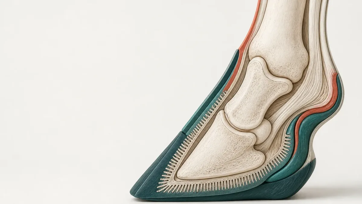
The complication with the gravest consequences strikes not the broken limb but the healthy one beside it. Inside the hoof a fine layer of tissue, the lamellar apparatus, interlocks the pedal bone with the hoof capsule. When that attachment fails, the pedal bone sinks or rotates: that is laminitis. When it appears in the limb opposite a painful injury, it is called supporting-limb laminitis.
A review from the group that supplies most of the experimental data describes the process in two steps. First the constant alternation between loading and unloading that normally goes with moving about is missing, and perfusion of the lamellae suffers. Then the extra load tips the already weakened tissue into acute laminitis.
Several measurements exist for the first step. In an experiment with thirteen Standardbreds, a marker of oxygen shortage reached a value of 196 in the lamellar tissue of the more heavily loaded limb, against 42 in the comparison horses; outside the loaded hooves nothing of the sort was measurable. In a randomised experiment with nine Standardbreds, measures of lamellar perfusion improved as soon as the load changed frequently: thirty minutes of walking was enough. An hour of one-sided standing, by contrast, changed nothing measurable, and an hour says nothing about several days.
Imaging published in 2026 in eight healthy horses found, during quiet standing, areas of the inner sole in all eight animals and areas of the lamellae in seven of eight that did not take up a marker of perfusion. Those areas were absent once the horses had been allowed to move beforehand. The authors state expressly that this does not establish a link with supporting-limb laminitis in affected horses.
Is it the load or the standing still?
Probably both, and standing still weighs more heavily than long assumed. In an experiment from 2025, restricted freedom of movement without any extra load was enough to multiply dying cells in the lamellar tissue: 45 instead of four per lamella. The group concludes from this that movement has to be preserved, not merely load avoided.
How common this complication really is
Two figures from the same world appear to contradict each other. In the caseload of a British practice with 65,327 registered horses, eleven cases of supporting-limb laminitis were found over nine years, that is 0.02 per cent. They appeared between four and a hundred days after the injury, at a median of fourteen and a half days, and close to three quarters of those affected were Thoroughbreds. The authors also record that the complication was not confined to horses that had stopped bearing any weight at all on the injured limb.
Look instead only at horses that really are sparing a limb, and the figure becomes large. In an analysis of 113 horses in casts, fourteen developed supporting-limb laminitis, that is twelve per cent. Two features went with it: body weight and the duration of the cast. Whether a fracture was present made no difference. Any painful one-sided lameness can lead to it.
The recovery phase: the fracture that happens under anaesthesia
The second point at which fracture treatment fails lies not in the stable but in the recovery box. After a general anaesthetic a horse gets up under its own power, often before it can judge the situation, and the freshly screwed limb immediately takes the full mass.
How much that step weighs is shown by a worldwide survey of 47,396 general anaesthetics from 93 centres in 28 countries, in which the Vetsuisse Faculty in Zurich also took part. Mortality within seven days was 1.2 per cent overall and 0.6 per cent for procedures other than colic, in both cases lower than in the preceding survey of 2002. Among the 227 deaths outside colic, fractures came first at 35.7 per cent, ahead of abdominal complications at 18.1 per cent. The authors record that these fractures arose predominantly during the recovery phase, and that healthy horses were among them.
That figure describes the anaesthetic risk of a mixed population, not the outlook for a horse with a freshly operated fracture. Centres reported voluntarily, which over-represents well equipped clinics and, if anything, understates true mortality.
Which fractures are repaired today
The claim that a broken leg means the end for a horse is, in that generality, out of date. What decides is not the name of the bone but the form of the break: whether it stays closed, whether it splits into few or many pieces, and whether it can be screwed together stably.
Standing surgery shows this most clearly. In an analysis of 245 racehorses operated on standing under sedation between 2007 and 2021 for a longitudinal fissure of the pastern or the cannon bone, 98 per cent left the clinic alive, and around three quarters raced again, at a median of 241 days after the procedure. Earnings per start, the proportion of winners and the proportion of placed horses did not differ meaningfully before and after surgery. These are two selected fracture forms at a specialist centre.
For pastern joint arthrodesis, a meta-analysis of 21 studies covering 458 horses sums up: 90 per cent survived, 65 per cent returned to their intended use, 12 per cent developed a wound infection. That separation is the real point. Surviving and being usable again are two different outcomes, and around twenty-five percentage points lie between them here.
Why mass, age and infection weigh more than the technique
The same operation on the same bone can succeed almost every time or fail almost every time depending on the patient. In a case series on complete diaphyseal fractures of the fore and hind cannon bone, three of ten adult horses survived, but ten of eleven foals. In open fractures the gap was sharper still: one of eight adults against six of seven foals. The authors name body weight, age and infection as the decisive quantities and mark a weight above 320 kilograms in their series as a risk factor.
Infection is no marginal problem here. In an analysis of 155 horses with internal fixation, 14.2 per cent developed a surgical site infection. The affected animals left the clinic alive twelve times less often than the rest. Local antibiotic delivery could not be shown to be protective in this dataset.
History counts too. An analysis of 115 post-mortem reports from Californian racehorses with a complete tibial fracture found a pre-existing stress fracture in most of them; 68 per cent of the breaks happened in training rather than in a race, and 73 per cent in two- and three-year-old horses. A meta-analysis of risk factors in flat racing puts together a frequency of around 1.2 catastrophic musculoskeletal injuries per 1,000 starts and names age, race class, going, previous injury and findings at the pre-race examination as repeatedly associated factors.
The comparison with people
In people, comparable bone tissue heals under entirely different conditions. A Cochrane review from 2024 brings together 53 studies with 4,489 adults after surgically treated ankle fracture. Early weight bearing within three weeks probably improved function slightly over the first six months, without raising the frequency of further procedures. The difference remained small, however, and did not consistently reach a clinically meaningful threshold.
What is taken for granted in that work is impossible in the horse. A person chooses the timing and the dose of their loading, walks on crutches, sits, lies down and takes a splint off. The horse can do none of this. The question of whether to load early or late therefore does not transfer between the species: here it is not a treatment question but a question of species.
And if people are not operated on either?
Then the figures move closer together. A meta-analysis of eighteen studies on non-operated hip fracture in frail older people found a mortality of 36 per cent at thirty days and 60 per cent at one year; at six months just under ten per cent were mobile again.
This comparison has a limit that has to be named. The non-operated people in that analysis were the frailest patients of all, so their mortality mixes the consequences of the missing operation with their starting condition. The direction still reads clearly: where load-bearing function cannot be restored, a fracture becomes a life-threatening situation in people too. The horse is no special case in this. It only reaches that point earlier and more often.
What follows from this, and what does not
No rule for an individual horse follows from this evidence. It explains why the weighing-up after a broken leg looks different from the one in human medicine, and it explains just as much why that weighing-up has shifted: selected fracture forms can now be treated with a high probability of success, others still cannot.
What the studies do not supply is a yardstick by which an outsider could judge whether a particular decision was right. Fracture form, age, weight, temperament, level of pain and the possibility of weeks of aftercare all enter into it together, and none of the publications cited here weighs them against one another.
Outcome map of the published series: what each figure says and what it precisely does not
Original analysis
For every series found in this search, the reported measure has been set beside its population, the study design added, and the fourth column notes which conclusion does not follow from that particular figure; the rows come from different populations and are not comparable with one another.
Situation
Population
Reported outcome
What does not follow
Design
Standing osteosynthesis, longitudinal fissure of the pastern or cannon bone
245 racehorses, one centre, 2007 to 2021
98 per cent left the clinic alive, 75 per cent raced again, at a median of 241 days
Nothing about open or shattered fractures: the fracture forms were pre-selected
Retrospective cohort
Pastern joint arthrodesis
458 horses from 21 studies
90 per cent survived, 65 per cent returned to their intended use
That survival and usability are the same thing: around 25 percentage points lie between them
Meta-analysis of retrospective series
Complete diaphyseal fracture of the fore or hind cannon bone
10 adult horses and 11 foals
3 of 10 adults and 10 of 11 foals survived
That the technique fails: body mass, age and infection separate the groups
Case series
Disruption of the suspensory apparatus, double arthrodesis
26 racehorses, three clinics
13 of 26 were usable at pasture without painkillers beyond six months
That the rest stayed free of complications: 77 per cent had short-term complications
Case series
Infection after internal fixation
155 horses, one centre, 2008 to 2016
14.2 per cent developed a surgical site infection
That infection merely delays healing: those affected left the clinic alive twelve times less often
Retrospective cohort
Supporting-limb laminitis after a cast
113 casted horses at a referral clinic
12 per cent developed laminitis in the supporting limb
That a fracture determines the risk: body weight and cast duration did
Retrospective cohort
Supporting-limb laminitis in a practice caseload
65,327 horses in a British practice, 2005 to 2013
11 cases, equal to 0.02 per cent
That the complication is equally rare when a limb is already injured: the denominator is the whole caseload
Cross-sectional
Deaths around general anaesthesia
47,396 anaesthetics, 93 centres, 28 countries
0.6 per cent mortality within seven days excluding colic, of which 35.7 per cent from fractures
That the same risk applies to a horse with a freshly operated fracture: the population is mixed
Prospective cohort
Limitations and uncertainty
There are no randomised treatment studies of fractures in the horse, and there will be none: nobody randomises a broken leg. Six of the twenty works used here reach the level of systematic review, meta-analysis or randomised experiment, and two of those concern people. The rest are case series, cohorts, case-control analyses and experimental models.
Four of the works on supporting-limb laminitis come from the same research group, three of them from the same model with healthy, pain-free horses over 92 hours. They do not confirm one another independently, and none of those horses actually developed laminitis.
The outcome figures on fracture care come almost without exception from single specialist referral clinics and from pre-selected fracture forms. They describe what was achieved there with those patients, not the outlook for any horse with any break.
A large part of the data comes from racing, predominantly from Thoroughbreds and Standardbreds in Britain and the United States. Comparable figures for leisure and sport horses in Switzerland could not be found in this search.
The frequency figures for supporting-limb laminitis rest on eleven and on fourteen cases respectively. Numbers that small carry no dependable proportions, and searching electronic clinical records probably understates the true frequency.
The two human works describe ankle and hip fractures, not the counterpart of a cannon bone break. They serve to compare the surrounding conditions, not individual bones, and no course of action for the horse follows from them.
Open questions
Can the perfusion deficit measured in the standing hoof of the healthy horse be demonstrated at all in patients with a genuinely injured limb?
How much movement, how often and in what form would be needed to protect the lamellar tissue of the supporting limb over weeks?
Why do some severely lame horses never develop supporting-limb laminitis while others do so after only two weeks?
Do improved recovery procedures after general anaesthesia measurably shift the share of fractures among the deaths, or does getting up remain the limiting step?
Frequently asked questions
Can a horse survive a broken leg?
Yes, and for certain fracture forms it is now the rule. Of 245 racehorses operated on standing under sedation for a longitudinal fissure of the pastern or the cannon bone, 98 per cent left the clinic alive and around three quarters raced again. For a complete diaphyseal fracture of a large bone shattered into many pieces the picture reverses: in one case series three of ten adult horses survived, but ten of eleven foals. The outcome hangs on the form of the break, on body weight, on age, and on whether the repair holds stably.
Why is a horse with a broken leg often put down?
Because the weighing-up rarely concerns the break alone. A horse cannot lie down for weeks, it shifts the load onto the opposite limb and puts that hoof at risk. After a general anaesthetic it has to get up under its own power, and in a worldwide survey fractures were the commonest cause of death outside colic at around 36 per cent. Add a fracture form that cannot be screwed together stably, and what remains is weeks of painful treatment with a poor outlook. That weighing-up is done by the attending practice together with the owner or keeper, not by a statistic.
What is supporting-limb laminitis?
A disease of the healthy limb, not the broken one. When one limb carries more weight for days to weeks because the other one hurts, the attachment between the pedal bone and the hoof capsule suffers inside the hoof. In experiments with healthy horses a marker of oxygen shortage rose markedly in the more heavily loaded limb, and restricted freedom of movement on its own already raised the number of dying cells in the lamellar tissue. In the caseload of a British practice the complication was rare in absolute terms at 0.02 per cent; after a cast, by contrast, it occurred in twelve of every hundred horses.
Does a horse's bone heal worse than a human bone?
No, and that is the commonest misconception around this question. The bone itself heals. What is missing are the conditions: a person can offload a fracture, walk on crutches, sit and lie down, and choose when to bear weight. A Cochrane review of surgically treated ankle fractures compares exactly such loading plans, and the horse has no such choice. Then there is the mass: at gallop, peak forces of 14.0 newtons per kilogram of body weight were measured on one foreleg, and even in quiet standing a single foreleg carries about a quarter of the body weight.
Why is the leg not simply put in a cast?
Because the cast itself carries a risk. In an analysis of 113 casted horses, twelve per cent developed supporting-limb laminitis, and two quantities went with it: body weight and the duration of the cast. Full-limb casts and those with transfixation pins loaded the limb more than half-limb casts. A cast also immobilises exactly the movement that, according to the available experiments, keeps perfusion going inside the hoof. For some fractures it is the right choice, but it is not a harmless interim measure.
Is it really so different in people?
In the care, yes; in the outcome, not always. As long as surgery is possible, the human outlook is incomparably better. Where load-bearing function is not restored, that changes: in a meta-analysis of eighteen studies on non-operated hip fractures in frail older people, mortality at one year was 60 per cent, and at six months just under ten per cent were mobile again. Those people were, however, the frailest patients of all, which is why the share attributable to the missing operation and the share attributable to their starting condition cannot be separated.
Sources
Self Davies ZT, Spence AJ, Wilson AM. Ground reaction forces of overground galloping in ridden Thoroughbred racehorses. The Journal of Experimental Biology, 2019 (Laboratory study | Horse)DOI 10.1242/jeb.204107 Direct force measurement in seven ridden Thoroughbreds at gallop: the peak vertical force reached 14.0 newtons per kilogram on the non-leading foreleg, and the load was split roughly evenly between the forehand and the hindquarters rather than in the often quoted sixty-to-forty ratio.
van Eps AW, Belknap JK, Schneider X, Stefanovski D. Lamellar perfusion and energy metabolism in a preferential weight bearing model. Equine Veterinary Journal, 2021 (Controlled trial | Horse)DOI 10.1111/evj.13356 In the model of an enforced offloading posture the observed foreleg carried a median of 38.7 instead of 27.3 per cent of body weight, and signs of oxygen shortage in the lamellar tissue rose only there: the authors read the reduced perfusion as a consequence of the raised mechanical load.
Engiles JB, Stefanovski D, van Eps A. Lamellar cell death and proliferation are associated with restricted ambulation and preferential weight bearing in a model relevant to supporting-limb laminitis. American Journal of Veterinary Research, 2025 (Controlled trial | Horse)DOI 10.2460/ajvr.24.09.0268 Restricted freedom of movement without any added load already led to cell death in the lamellar tissue (45 against 4 per lamella), which is why the authors name preserving movement, and not load reduction alone, as a possible protective strategy.
Medina-Torres CE, Underwood C, Pollitt CC, Castro-Olivera EM. The effect of weightbearing and limb load cycling on equine lamellar perfusion and energy metabolism measured using tissue microdialysis. Equine Veterinary Journal, 2016 (Randomised trial | Horse)DOI 10.1111/evj.12377 In a randomised experiment with nine Standardbreds, frequent load cycling and thirty minutes of walking improved measures of lamellar perfusion, whereas an hour of one-sided standing and sedation changed nothing measurable.
Skelton G, Wulster-Bills K, Ciamillo S, Anishchenko S. Positron emission tomography using 18F-fluorodeoxyglucose reveals digital perfusion deficits associated with continuous weight-bearing in healthy standing horses. American Journal of Veterinary Research, 2026 (Laboratory study | Horse)DOI 10.2460/ajvr.25.07.0268 In eight healthy horses standing quietly, imaging showed areas of the inner sole and of the lamellae without tracer uptake that resolved with movement; the authors stress expressly that the significance for supporting-limb laminitis in patients remains open.
Burns TA, Watts MR, Belknap JK, van Eps AW. Digital lamellar inflammatory signaling in an experimental model of equine preferential weight bearing. Journal of Veterinary Internal Medicine, 2023 (Randomised trial | Horse)DOI 10.1111/jvim.16662 In the same model of an enforced offloading posture, inflammatory signalling in the lamellar tissue of the loaded limb was raised; the authors treat these pathways as possibly important, in the conditional, and rank them neither above nor below reduced perfusion as a cause.
van Eps A, Engiles J, Galantino-Homer H. Supporting Limb Laminitis. Veterinary Clinics of North America: Equine Practice, 2021 (Other | Horse)DOI 10.1016/j.cveq.2021.08.002 Narrative review describing the process in two steps: first the missing alternation between loading and unloading disturbs perfusion of the lamellae, then the extra load tips the weakened tissue into acute laminitis; preserving load cycling counts for the authors as the central preventive measure.
Wylie CE, Newton JR, Bathe AP, Payne RJ. Prevalence of supporting limb laminitis in a UK equine practice and referral hospital setting between 2005 and 2013: implications for future epidemiological studies. The Veterinary Record, 2015 (Cross-sectional study | Horse)DOI 10.1136/vr.102426 In a caseload of 65,327 horses only eleven cases were found over nine years, that is 0.02 per cent, occurring between four and a hundred days after the injury; the authors conclude above all that future risk factor studies will founder on the numbers available.
Virgin JE, Goodrich LR, Baxter GM, Rao S. Incidence of support limb laminitis in horses treated with half limb, full limb or transfixation pin casts: a retrospective study of 113 horses (2000-2009). Equine Veterinary Journal Supplement, 2011 (Cohort study | Horse)DOI 10.1111/j.2042-3306.2011.00491.x Of 113 casted horses, 14, that is 12 per cent, developed supporting-limb laminitis; only body weight and cast duration were significantly associated, not the presence of a fracture, the limb affected or the breed.
Gozalo-Marcilla M, Redondo JI, Bettschart-Wolfensberger R, Domenech L. The Confidential Enquiry into Perioperative Equine Fatalities: phase 4 (CEPEF4) - a worldwide observational, prospective, multicentre cohort study in 2025. Veterinary Anaesthesia and Analgesia, 2025 (Cohort study | Horse)DOI 10.1016/j.vaa.2025.06.005 Across 47,396 general anaesthetics from 93 centres in 28 countries, mortality within seven days was 1.2 per cent overall and 0.6 per cent excluding colic; among the 227 deaths outside colic, fractures came first at 35.7 per cent, predominantly during the recovery phase.
Hitchens PL, Morrice-West AV, Stevenson MA, Whitton RC. Meta-analysis of risk factors for racehorse catastrophic musculoskeletal injury in flat racing. The Veterinary Journal, 2019 (Meta-analysis | Horse)DOI 10.1016/j.tvjl.2018.11.014 From 21 incidence studies and 65 risk factor studies comes a pooled frequency of 1.17 catastrophic musculoskeletal injuries per 1,000 starts in flat racing, with age, race class, going, previous injury and pre-race examination findings as consistently associated factors.
Samol MA, Uzal FA, Hill AE, Arthur RM. Characteristics of complete tibial fractures in California racehorses. Equine Veterinary Journal, 2021 (Case-control study | Horse)DOI 10.1111/evj.13375 Among 115 post-mortem findings, complete tibial fractures were predominantly associated with a stress fracture arising early in the career; 68 per cent happened in training, 73 per cent in two- and three-year-old horses, and the affected animals had fewer starts and fewer training kilometres than the comparison horses.
Colgate VA, Robinson N, Barnett TP, Bathe AP. Outcome and racing performance following standing fracture repair in 245 horses. Equine Veterinary Journal, 2024 (Cohort study | Horse)DOI 10.1111/evj.14016 In 245 racehorses operated on standing, survival to discharge was 98 per cent and return to racing 75.1 per cent after a median of 241 days, with no meaningful difference in performance before and after the procedure; the cases involved selected fracture forms.
de Souza AF, Paretsis NF, De Zoppa ALDV. Proximal Interphalangeal Arthrodesis in Horses: A Meta-Analysis of Retrospective Studies. Journal of Equine Veterinary Science, 2023 (Meta-analysis | Horse)DOI 10.1016/j.jevs.2023.104226 Across 21 studies with 458 horses, survival was 90 per cent, return to intended use 65 per cent and wound infection 12 per cent; the authors call the procedure safe but consider usability in need of improvement, particularly after fractures.
Bischofberger AS, Fürst A, Auer J, Lischer C. Surgical management of complete diaphyseal third metacarpal and metatarsal bone fractures: clinical outcome in 10 mature horses and 11 foals. Equine Veterinary Journal, 2009 (Case series | Horse)DOI 10.2746/042516409x389388 With complete diaphyseal fractures, 3 of 10 adult horses survived against 10 of 11 foals, and the authors name age, a body weight above 320 kilograms, unstable fixation and infection as the decisive quantities behind failure.
Curtiss AL, Stefanovski D, Richardson DW. Surgical site infection associated with equine orthopedic internal fixation: 155 cases (2008-2016). Veterinary Surgery, 2019 (Cohort study | Horse)DOI 10.1111/vsu.13216 Of 155 horses with internal fixation, 14.2 per cent developed a surgical site infection, and the affected animals left the clinic alive twelve times less often; no protective effect of local antibiotic delivery could be shown.
Orozco Lopez D, Garcia-Lopez JM, Carpenter R, Bras JJ. Treatment of traumatic disruption of the suspensory apparatus in Thoroughbred racehorses at risk of proximal interphalangeal joint subluxation using a locking compression-distal femur plate for double arthrodesis. Veterinary Surgery, 2025 (Case series | Horse)DOI 10.1111/vsu.14219 In 26 racehorses the double arthrodesis prevented every subluxation of the pastern joint, yet 76.9 per cent had short-term complications and only half were usable at pasture without painkillers beyond six months; supporting-limb laminitis is among the complications named.
Bowers K, Weinhandl JT, Anderson DE. A review of equine tibial fractures. Equine Veterinary Journal, 2023 (Other | Horse)DOI 10.1111/evj.13599 Narrative review that ties the prognosis to the form of the break: complete tibial fractures in the adult horse offer few treatment routes and a comparatively high mortality, whereas stress fractures and minimally displaced incomplete fractures usually heal well without surgery.
Lewis SR, Pritchard MW, Parker R, Searle HKC. Rehabilitation for ankle fractures in adults. Cochrane Database of Systematic Reviews, 2024 (Meta-analysis | Human)DOI 10.1002/14651858.CD005595.pub4 Across 53 studies with 4,489 adults, early weight bearing after surgically treated ankle fracture probably improved function slightly over the first six months without raising the number of further procedures, the difference remaining small and blinding having been impossible in every study.
Loggers SAI, Van Lieshout EMM, Joosse P, Verhofstad MHJ. Prognosis of nonoperative treatment in elderly patients with a hip fracture: A systematic review and meta-analysis. Injury, 2020 (Meta-analysis | Human)DOI 10.1016/j.injury.2020.08.027 From 18 studies comes, for non-operated hip fracture in frail older people, a mortality of 36 per cent at 30 days and 60 per cent at one year, with just under 10 per cent of patients still mobile at six months, this group comprising the frailest patients of all.
ForschungPferd (2026). Why a broken leg ends differently in a horse than in a person. ForschungPferd, English. https://forschungpferd.ch/en/comparative-health/equine-fracture-outcome-comparison/